
ITGB3 & Multiple Sclerosis: When Doctoral Research Becomes an Animation
Scientific Advisor: Dr. Giselle Vela, PhD — Molecular Biology in Medicine
Project type: Scientific visualization / Mechanism of action animation
Sector: Medical & neuroscience research
Reference: https://www.mdpi.com/1422-0067/26/24/12094
The Challenge
Multiple sclerosis affects nearly 3 million people worldwide, with a paradox at its core: women develop it three times more often than men, but men experience more severe neurological damage. Understanding why requires going deeper than clinical observation.
Dr. Giselle Vela's doctoral research focused on a specific question: what role do pleiotrophin (PTN) receptors play in peripheral T-lymphocytes in MS? PTN is a cytokine significantly elevated in relapsing-remitting MS patients — but its receptor interactions in peripheral immune cells remained poorly understood.
Her team's key finding: significant overexpression of integrin subunit beta-3 (ITGB3) mRNA in RRMS patients vs. healthy controls, with molecular docking evidence suggesting PTN binds ITGB3 with higher affinity than fibrinogen, its canonical endogenous ligand.
The mechanism — spanning cytokine signaling, receptor expression, and immune cell trafficking across the blood-brain barrier — was invisible to anyone outside the field. That's the problem FIS was brought in to solve.
The Science Behind the Animation
To produce this animation, FIS worked directly from Dr. Vela's doctoral research. Every sequence in the animation corresponds to a mechanism documented in her work. Here is what the animation shows, scene by scene:
The blood-brain barrier under normal conditions The central nervous system is shielded by one of the body's most selective structures. The animation opens inside a blood vessel, establishing the environment before showing what happens when that environment fails.
Epstein-Barr virus and T-cell activation In multiple sclerosis, the EBV infection plays a key role. Antigen-presenting cells (APCs) activate CD4+ T-lymphocytes that have been primed by viral antigens, driving their differentiation into pro-inflammatory phenotypes — TH1 and TH17. The animation visualizes this interaction at the molecular level inside the vascular environment.
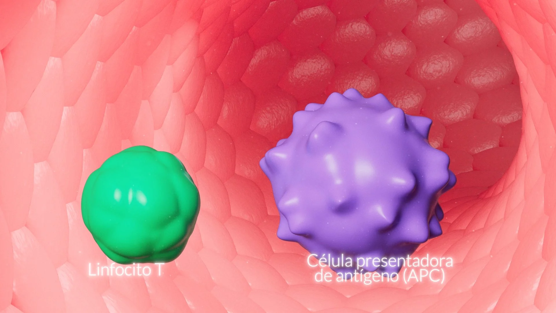
T-cell trafficking through the bloodstream Activated T-cells enter circulation. The animation follows a single lymphocyte through the vessel, surrounded by red blood cells, establishing scale and biological context before the next critical event.
Adhesion to the vascular endothelium When these cells reach the blood-brain barrier, they interact with the endothelium through adhesion molecules — selectins, ICAM-1, and VCAM-1. The animation shows the lymphocyte decelerating, rolling, and adhering to the vessel wall. This is the moment before invasion.
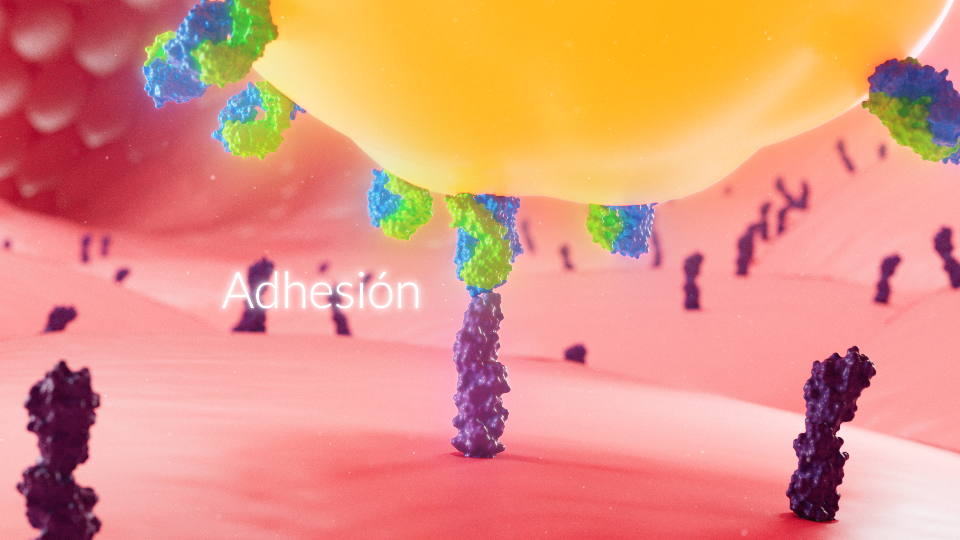
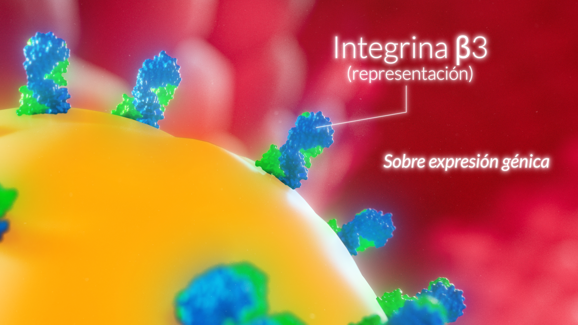
The ITGB3 finding — the key discovery The central finding of Dr. Vela's research: the overexpression of ITGB3 in these T-cells. This integrin, expressed on the surface of the lymphocyte, is visualized at the molecular level — its structure, its binding, and how its overexpression may enhance adhesion to the endothelium and facilitate diapedesis into the central nervous system.
Diapedesis — crossing the barrier The T-cell migrates through the endothelial wall. The animation renders this extravasation sequence with anatomical precision, showing the mechanical process of a cell breaching a biological barrier.
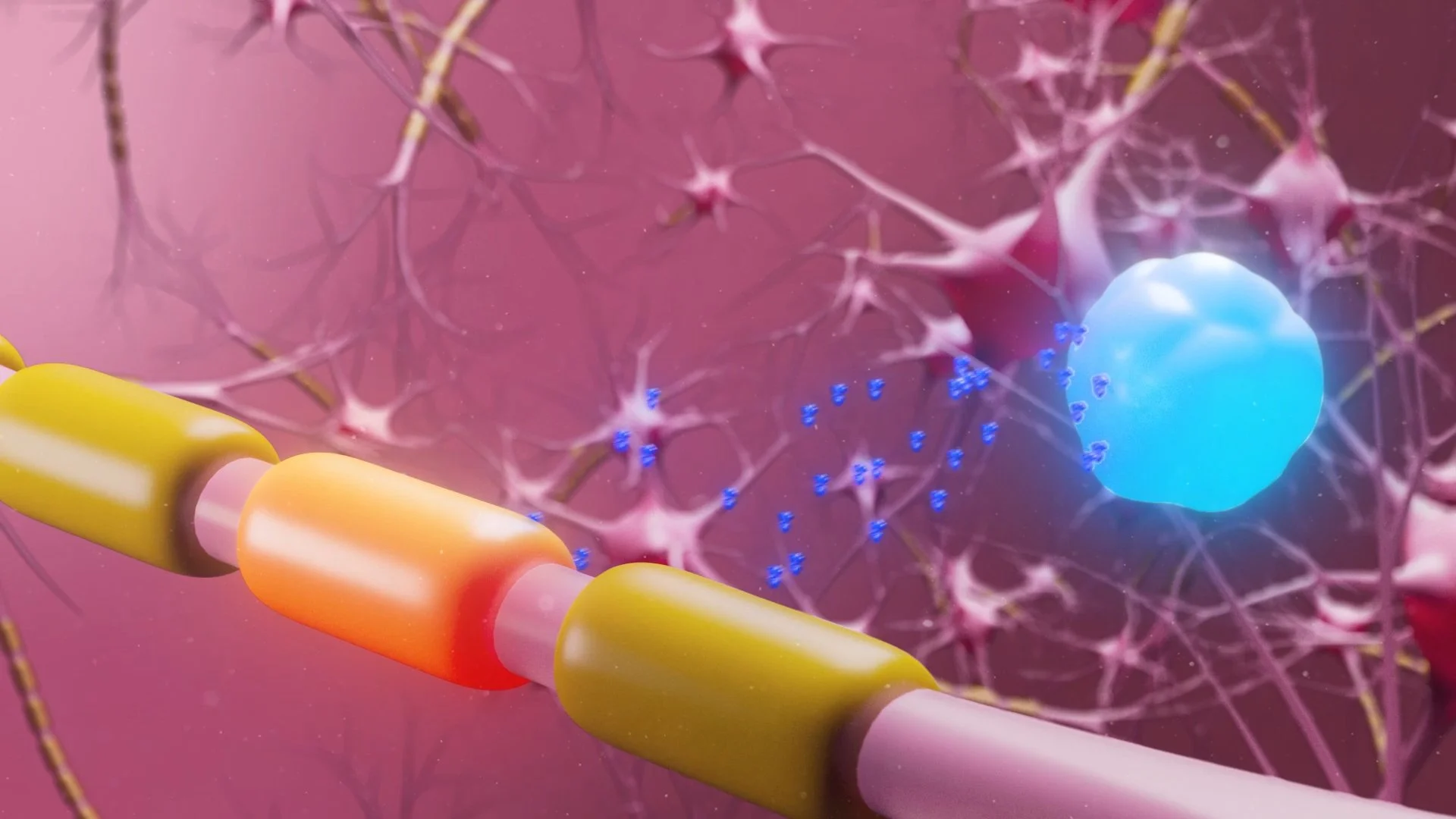
Molecular mimicry and myelin attack Once inside the CNS, the mechanism of molecular mimicry is shown: structural similarities between an EBV protein and myelin basic protein cause the immune system to misidentify and attack the myelin sheath. The animation shows the myelin degrading — the physical basis of MS symptoms.
Neuronal damage and the breakdown of neural communication The consequence: demyelinated axons, disrupted neural signaling, and the cascade of symptoms that define the disease. The animation closes on damaged neural tissue, connecting molecular events to clinical reality.
Regulatory T-cells — the failed counterbalance The final sequence shows regulatory T-cells releasing anti-inflammatory cytokines — the body's attempt at control — and how this equilibrium breaks down during active disease.
The Production Approach
FIS built every element in this animation from scientific reference, not from stock assets. The cellular morphology, the molecular structures, the vascular environment — each was modeled to reflect what Dr. Vela's research describes, not what a generic biology illustration might approximate.
The color system was designed for clarity: each cell type and molecular actor is visually distinct and consistent across the full 120 seconds. The camera work prioritizes comprehension — wide establishing shots that give context, macro sequences that reveal mechanism, transitions that preserve spatial logic between scales.
The result is an animation that a neurologist and a non-specialist can watch together and both understand.
The Result
A 120-second scientific animation that translates the full mechanism of ITGB3-mediated T-cell trafficking in multiple sclerosis — from viral activation to neural damage — into a visual narrative grounded in doctoral research.
This is the kind of work FIS was built to do: not decorative science, but functional visualization that makes complex biological mechanisms impossible to misunderstand.
What This Means for Your Project
If your organization is working with complex biological mechanisms, clinical findings, or research that needs to communicate beyond your immediate field — this is exactly the problem FIS solves.
We work with biotech and pharmaceutical teams to produce scientific animations for investor presentations, regulatory submissions, grant proposals, and patient education. Every project starts with the science. The visuals follow.
Frame Imagery Solutions specializes in medical and scientific visualization for biotech, pharma, and research institutions. All projects are produced in close collaboration with the scientific teams behind the research.